



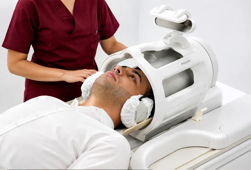
A 3 Tesla MRI is a magnetic resonance scanner with a 3 Tesla magnetic field strength, designed to produce high-resolution, sharp images. The magnet is the foundation of any MRI system, and a 3 Tesla field strengthens doctors' diagnostic capabilities while giving patients a faster, more detailed examination. NPİSTANBUL Hospital uses a wide-bore (70 cm) 3 Tesla MRI scanner, a device found in only a few centres in Türkiye, to combine high image quality with patient comfort.
Advantages of the 3 Tesla MRI at NPİSTANBUL
- A wider, more comfortable scanner: the roomy interior makes it easier for patients to stay comfortable and cooperate during the scan, and makes MRI possible for people with claustrophobia or severe obesity.
- Less noise: loud noise during the examination is one of the main sources of discomfort in 3T scanners, and some patients find it very distressing. This device uses a noise-reduction technique that can cut the sound the patient hears by up to 40 percent.
- Music and video: a music system and video broadcast help patients pass the time pleasantly during the scan.
- High image quality in every scan: software corrects image distortion caused by movement and by MR-compatible metal prostheses and implants in the body.
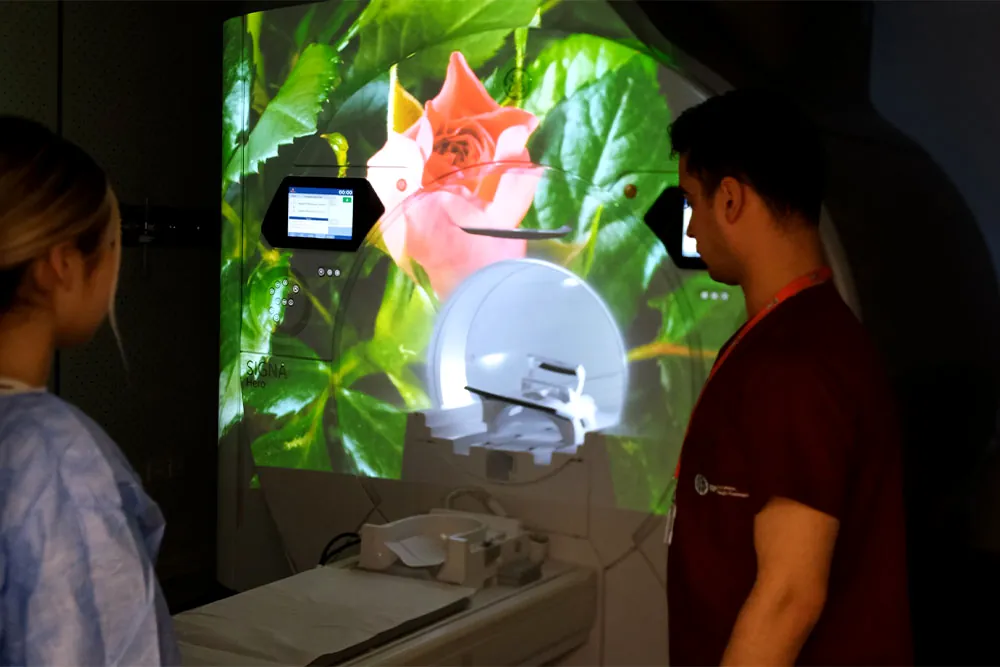
- A child-friendly experience: a dedicated approach helps children feel calmer and more at ease, especially those with claustrophobia or anxiety. Ambient effects inside and outside the scanner, special images and music are used to distract and soothe them. For example, coloured lights or pleasant landscape images can draw a child's attention away from the scan, and soft music can help them relax.
- Sedation when needed: for people with special needs, scans under sedation anaesthesia are carried out safely.
What to expect during the scan
- Remove all metal: nothing metallic, such as necklaces, earrings, watches or rings, may be worn. The team will ask you to take off any metal items first.
- Tell us about implants and pregnancy: before the scan, let the team know if you have a pacemaker or defibrillator, a neurostimulator, a cochlear implant, aneurysm clips, a medication pump, metal fragments in your body, or tattoos or permanent make-up, and whether you are or may be pregnant. Some implants are not safe in a 3 Tesla scanner, so each one is checked before you enter the room.
- Positioning: you lie on the scanner table, and the team shows you how to lie depending on the area being imaged.
- Headphones: you hear and follow instructions through headphones during the scan, and they also reduce the noise of the machine.
- Call button: you hold a button that lets you alert the team quickly if there is any problem.
- Duration: scan time depends on the area being imaged and the type of condition being investigated.
What 3 Tesla MRI is used for
3 Tesla MRI can image organs and systems throughout the body. It is used to detect or assess:
- Brain tumours
- Epilepsy
- Spinal cord diseases
- Neurological disorders such as migraine
- Ear disorders
- Jaw problems
- Spine diseases
- Prostate diseases
- Breast diseases
- Joints and connective tissue, such as the shoulders and knees
- Many bone-related problems
- The coronary vessels that supply the heart
- Whole-body imaging
When and why is MRI ordered?
MRI is one of the advanced radiological examinations that play a leading role in diagnosing disease. It is requested when other imaging methods cannot establish a diagnosis, or when findings do not explain the patient's clinical condition.
MRI is also among the first tests requested for many organs that plain X-rays cannot show, such as the brain. Computed tomography gives better images of bone, while MRI is better at showing soft tissue.
Advanced imaging techniques
- Advanced MRI imaging
- Virtual colonoscopy
- MR enterography
- Multiparametric prostate MRI
- MR fistulography
- Functional MRI
- MR spectroscopy (multivoxel, single-voxel and 3D voxel)
- Diffusion tensor imaging (DTI)
- MR perfusion
- MR cisternography
- MR arthrography
- Cartilage T2 mapping
In addition, the Radiology Clinic at NPİSTANBUL Hospital uses the latest computer software to produce colour maps and to examine images from different angles.
Used to diagnose
Used in the assessment of
- Brain and spine surgery Can be part of the assessment
- Depression In selected cases, to rule out other causes
- Schizophrenia and psychosis In selected cases, to rule out other causes
- Parkinson’s disease and movement disorders In selected cases, to rule out other causes
Programmes that include it
- Memory and neurological assessment Expected stay: 2–3 days
Often combined with
- Procedure Neuronavigation-guided surgery Surgery guided by real-time 3D imaging to reach the target precisely and protect healthy brain tissue.